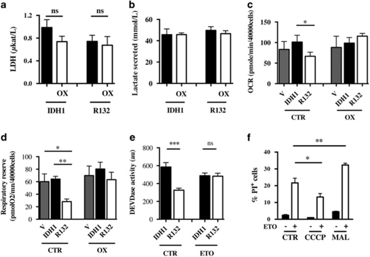
Figure 6.
Lactate deshydrogenase activity (a) and lactate production (b) of U251 cells expressing IDH1 isoforms. Cells were treated or not with 3 mM oxamate (OX) for 48 h. (c) Basal oxygen consumption was determined by measuring OCR as in Figure 4c in cells expressing IDH1 isoforms treated or not with 3 mM oxamate for 7 days. (d) Mitochondrial respiratory reserve was determined as in Figure 4e in cells treated or not with 3 mM oxamate for 7 days. (e) Caspase 3 activation was determined using a DEVDase activity assay 24 h after ETO-induced apoptosis in U251 cells treated or not for 7 days with 3 mM oxamate. (f) The number of dead cells was measured 24 h after a concomitant treatment with CCCP (1 μM) or malate (3 mM) and ETO (50 μg/ml). The number of dead cells was determined by FACS after propidium iodide (1 μg/ml) staining. Results are expressed as the mean±S.E.M. of three experiments performed in triplicate

