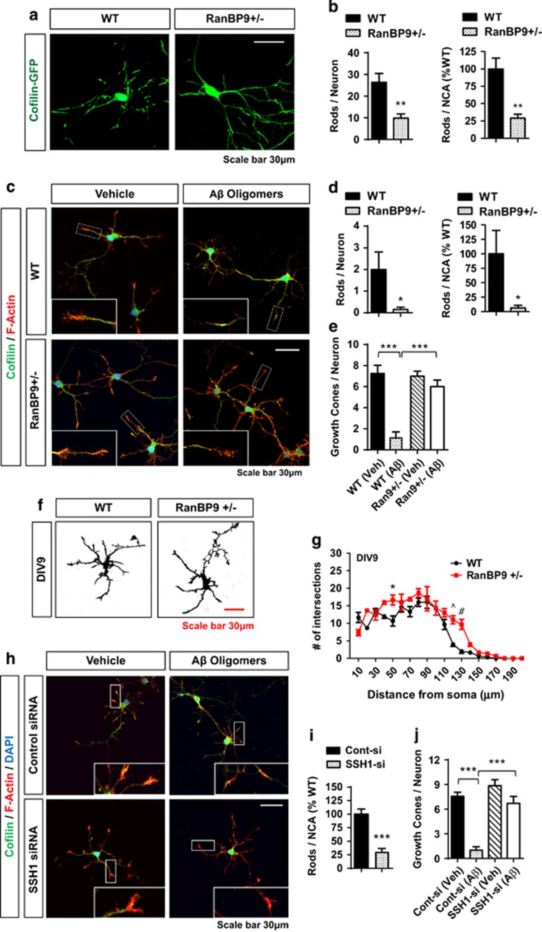
Figure 2.
RanBP9 mediates cofilin–actin rod formation and AβO-induced collapse of growth cones in primary hippocampal neurons. (a and b) DIV8 hippocampal neurons from WT and RanBP9+/− mice transiently transfected with cofilin-GFP and treated with hydrogen peroxide (1 μM) for 30 min and subjected to F-actin staining (Rhodamine-phalloidin). Representative images showing marked reduction in cofilin-GFP rods in RanBP9+/− neurons, despite strong GFP-cofilin fluorescence. (b) Quantitation of cofilin-GFP rods per neuron (t-test, **P=0.0029, n=6 replicates from three pups per genotype) or per NCA (t-test, **P=0.0009, n=6 replicates from three pups per genotype) after treatment of hydrogen peroxide (1 μM) for 30 min. NCA derived from area covered by saturated F-actin stain. (c-e) DIV8 hippocampal neurons from WT and RanBP9+/− mice treated with or without Aβ1-42O for 24 h and subjected to staining for F-actin (Rhodamine-phalloidin) and cofilin. (c) Representative images showing no cofilin rods observed without Aβ1-42O treatment but 16.3% and 6.9% of WT and RanBP9+/− neurons, respectively, contain cofilin rods. Also note the near absence of F-actin containing growth cones in WT neurons treated with Aβ42O but not in RanBP9+/− neurons. (d) Quantitation of Cofilin rods per neuron (t-test, *P=0.0329, n=6 replicates) or per NCA (t-test, *P=0.030, n=6 replicates from three pups per genotype). (e) Quantitation of F-actin containing growth cones in WT and RanBP9+/− (Ran9+/−) neurons with or without Aβ42O treatment (ANOVA, post-hoc Tukey, ***P<0.0001, n=8 replicates from four pups per genotype). (f and g) Scholl analysis of neurite arborization/elongation in WT and RanBP9+/− hippocampal neurons on DIV9. (f) Representative images of saturated F-actin stain. (g) Quantitation of neurite intersections on concentric circles from the soma in 10μm increments (10–200 μm) (two-way ANOVA, post-hoc Bonferroni, *P<0.05, ^P<0.005, #P<0.0005, n=3 replicates from two mice per genotype). Error bars represent S.E.M. in graphs. (h-j) DIV8 hippocampal neurons from WT mice with or without control or SSH1 siRNA transfection treated with or without Aβ42O for 24 h and subjected to staining for F-actin (Rhodamine-phalloidin) and cofilin. (h) Representative images showing cofilin and F-actin stains. (i) Quantitation of cofilin rods per NCA (t-test, ***P<0.0001, n=6 replicates from three pups per genotype). (j) Quantitation of F-actin containing growth cones in neurons transfected with control or SSH1 siRNA with or without Aβ42O treatment (ANOVA, post-hoc Tukey, ***P<0.0001, n=8 replicates from four pups per genotype)

