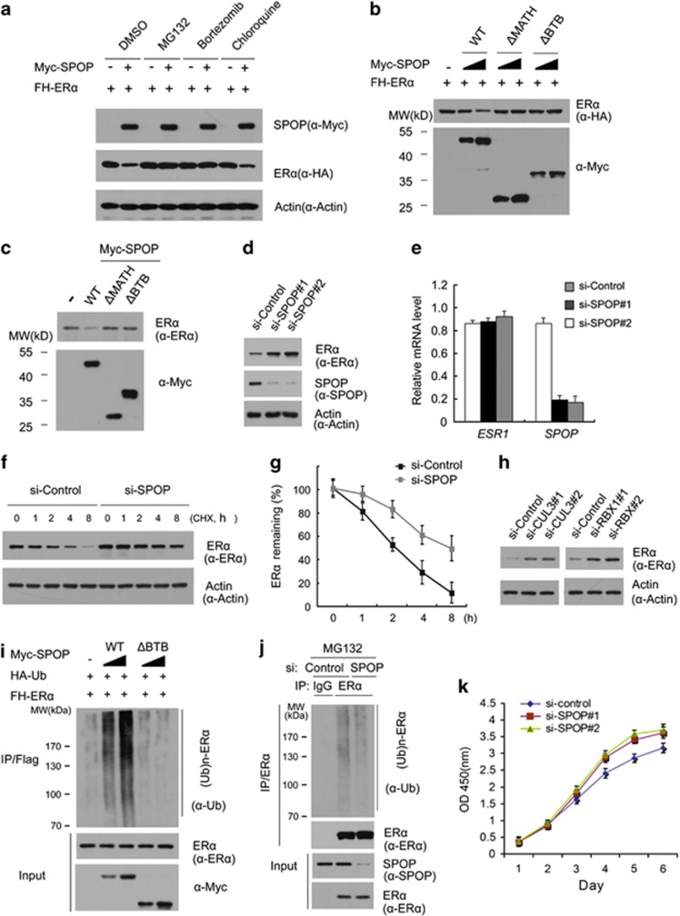
Figure 2.
The SPOP-CUL3-RBX1 ubiquitin ligase complex targets ERα for ubiquitination and degradation. (a) SPOP regulates ERα protein levels through the proteasome pathway. The 293T cells were transfected with FH-ERα in combination with or without the Myc-SPOP constructs. After 24 h, cells were treated with MG132 (20 μM), Bortezomib (200 nM), chloroquine (100 mM), or DMSO for 4 h before cell lysates were prepared for WB analyzes. Actin, a loading control. (b) The BTB and MATH domains in SPOP are essential for SPOP-mediated degradation of ERα. FH-ERα and different amounts of Myc-SPOP-WT or deletion mutants (ΔMATH, ΔBTB) constructs were transfected into 293T cells. After 24 h, cell lysates were prepared for WB analyzes. (c) SPOP regulates endogenous ERα protein levels. Ishikawa cells were transfected with Myc-SPOP-WT, or deletion mutants (ΔMATH, ΔBTB) constructs. After 24 h, cell lysates were prepared for WB analyzes. (d) Knockdown of SPOP increases endogenous ERα protein levels. Ishikawa cells were transfected with control or two SPOP-specific siRNAs. After 48 h, cell lysates were prepared for WB analyzes. (e) Quantitative RT-PCR measurement of the mRNA levels of SPOP and ESR1 in SPOP-knockdown Ishikawa cells. The mRNA level of GAPDH was used for normalization. The mean values (S.D.) of three independent experiments are shown. (f,g) Knockdown of SPOP prolongs ERα protein half-life. Ishikawa cells were transfected with control or SPOP-specific siRNA. After 48 h, cells were chased with 30 μM cycloheximide (CHX). At the indicated time points, cell lysates were prepared for WB analyzes. (f) At each time point, the intensity of ERα was first normalized to the intensity of Actin (loading control) and then to the value of the 0-h time point (g). The mean values (S.D.) of three independent experiments are shown. (h) Knockdown of RBX1 or CUL3 increases endogenous ERα protein levels. Ishikawa cells were transfected with control siRNA or siRNAs for RBX1 or CUL3. After 48 h, cell lysates were prepared for WB analyzes. (i) SPOP promotes ERα polyubiquitination in vivo. FH-ERα, HA-Ub, and Myc-SPOP-WT or ΔBTB mutant constructs were co-transfected into 293T cells. After 24 h, cells were treated with 20 μM MG132 for 4 h. ERα proteins were immunoprecipitated with anti-FLAG antibody and resolved by SDS/PAGE. The ubiquitinated forms of ERα were analyzed by WB with anti-Ub antibody. (j) Knockdown of SPOP decreases ubiquitination of endogenous ERα. Ishiwaka cells were transfected control or SPOP-specific siRNA. After 48 h, cells were treated with 20 μM MG132 for 4 h and then the same procedure was performed as i. (k) Knockdown of SPOP promotes Ishikawa cells growth. Ishikawa cells were transfected with control or two SPOP-specific siRNAs. After 48 h, the cell growth was measured by CCK-8 assay at indicated days. The mean values (S.D.) of three independent experiments are shown

