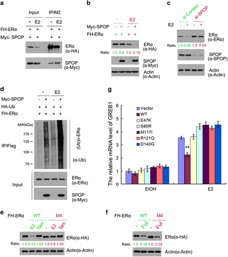
Figure 5.
Estrogen potentiates SPOP-mediated degradation of ERα. (a) Estrogen enhances the SPOP-ERα interaction. FH-ERα and Myc-SPOP constructs were co-transfected into 293T cells. After 24 h, cells were treated with the vehicle ethanol (EtOH,−) or 10 nM 17β-estradiol (E2) for 4 h before cell lysates were prepared for co-IP and WB analyzes. (b) Estrogen enhances SPOP-mediated ERα degradation. The 293T cells were transfected with the indicated constructs. A small amount of Myc-SPOP constructs was used in transfection. After 24 h, cells were treated with the vehicle ethanol (EtOH) or 10 nM 17β-estradiol (E2) for 4 h before cells lysates were prepared for WB analyzes. The density of ERα was determined by normalizing to actin (loading control) first and then to the normalized value in mock-treated cells. (c) Knockdown of SPOP attenuates estrogen-induced degradation of ERα. Ishikawa cells were transfected with control or SPOP-specific siRNA. After 48 h, cells were then treated with the vehicle ethanol (EtOH,−) or 10 nM 17β-estradiol (E2) for 4 h before cell lysates were prepared for WB analyzes. (d) Estrogen potentiates SPOP-induced polyubiquitination of ERα. The 293T cells were transfected with the indicated constructs. After 24 h, cells were treated with the vehicle ethanol (EtOH,−) or 10 nM 17β-estradiol (E2). Cells were then treated with MG132 for 4 h before cell lysates were prepared for IP and WB analyzes. (e) Ishikawa cells lines that stably transfected with control, SPOP-WT or SPOP mutants constructs were treated with 10 nM 17β-estradiol (E2) for 24 h. The mRNA level of ERα target gene GREB1 was measured by qRT-PCR. The mRNA level of GAPDH was used for normalization. The mean values (S.D.) of three independent experiments are shown. **indicates statistical significance (**P<0.01). (f, g) Differential effects of estrogen on the protein level of ERα-WT and the SPOP degradation-resistant mutant (ERα-M4). The 293T cells were transfected with FH- ERα-WT or M4 mutant construct. After 24 h, cells were treated with vehicle ethanol (EtOH,−), 10 nM 17β-estradiol (E2), 10 nM Tamoxifen (Tam), and 10 nM Fulvestrant (Ful) for 4 h before cell lysates were prepared for WB analyzes

