Figure 3.
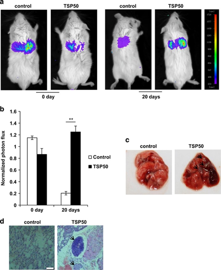
Overexpression of TSP50 promoted lung metastasis. (a) 2 × 10⁶ MCF-10A cells stably expressing control and TSP50 were injected through the tail vein of BALB/C mice. Mice were anaesthetized and injected with D-luciferin followed by bioluminescence imaging analysis of the lung metastasis as described in materials and methods section. (b) Quantification of bioluminescent imaging data. **P<0.01. (c) Appearance of the lungs from mice injected intravenously with control and TSP50-expressing cells. (d) Hematoxylin-eosin staining assay represent the metastases in the lungs. The arrow points out the metastasis nodules. Scale bar, 200 μm

