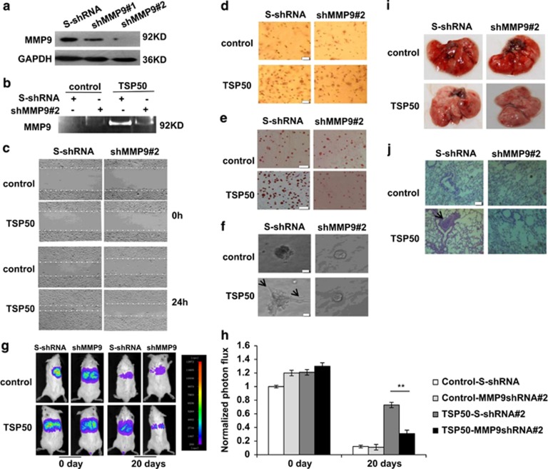
Figure 6.
MMP9 is required for TSP50-mediated cell migration, invasion and metastasis. (a) western blotting assay of MMP9 expression in control and TSP50-expressing CHO cells transfected with scrambled shRNA and TSP50 shRNA vectors. (b) Knockdown of MMP9 decreased MMP9 activities by gelatin zymography. (c) Knockdown of MMP9 inhibited TSP50-induced cell migration. Scale bar, 100 μm. (d) Knockdown of MMP9 inhibited TSP50-induced cell invasion. Scale bar, 50 μm. (e) Knockdown of MMP9 inhibited TSP50-induced cell adhesion. Scale bar, 150 μm. (f) Knockdown of MMP9 changed the morphology of TSP50-expressing CHO cells. The arrow points out the filopodium. Scale bar, 50 μm. (g) The effect of MMP9 knockdown on lung metastasis of TSP50-expressing cells was examined by bioluminescence imaging analysis. (h) Quantification of bioluminescent imaging data. **P<0.01. (i) Appearance of the lungs from mice injected intravenously with control and MMP9-silencing cells. (j) Hematoxylin and eosin staining assay represent the metastases in the lungs. The arrow points out the metastasis nodules. Scale bar, 200 μm

