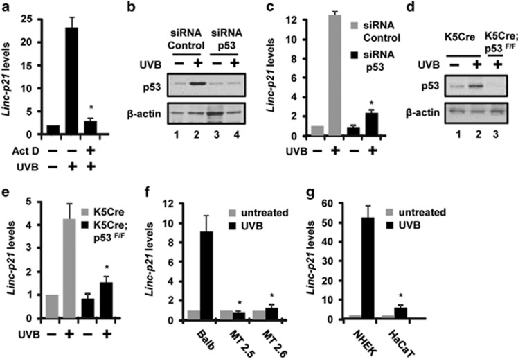
Figure 2.
LincRNA-p21 is regulated at the transcriptional level and is p53-dependent in mouse and human keratinocytes and mouse skin in vivo. (a) Balb/MK2 keratinocytes were treated with actinomycin D, exposed to 10 mJ/cm2 UVB, collected 12 h later and lincRNA-p21 transcripts measured. (b) Balb/MK2 cells were transfected with siRNA to p53 or control siRNA, and 48 h post transfection, cells were exposed to 10 mJ/cm2 UVB, collected 12 h later and p53 protein levels were measured by immunoblot analysis. (c) Balb/MK2 cells were transfected with siRNA to p53 or control siRNA, and 48 h post transfection, cells were exposed to 10 mJ/cm2 UVB, collected 12 h later and lincRNA-p21 transcripts measured. (d) K5Cre+/tg and K5Cre+/tg;p53flox/flox SKH-1 mice were treated with 200 mJ/cm2 UVB and epidermis collected 9 h later and p53 protein levels were measured by immunoblot analysis. (e) K5Cre+/tg and K5Cre+/tg;p53flox/flox mice were treated with 200 mJ/cm2 UVB and epidermis collected 9 h later and lincRNA-p21 transcripts measured. (f) MT2.5, MT2.6 and Balb/MK2 cells were exposed to 10 mJ/cm2 UVB and collected 8 h later. (g) NHEK and HaCaT cells were exposed to 10 mJ/cm2 UVB and collected 12 h later. LincRNA-p21 transcript levels were determined by TaqMan real-time PCR. Expression of lincRNA-p21 was normalized to β-actin. Data expressed in a, c, e, f and g as the mean ±S.D. N ≥3, *P<0.05 significantly different compared to UVB exposed control as determined by the student t-test

