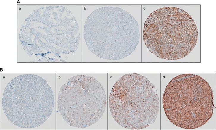
Figure 3.
Immunohistochemical analysis of STAT1 in HGSC. (A) The STAT1 antibody (Abcam no. ab2415, polyclonal rabbit anti-human STAT1) optimisation by IHC was performed in (a) normal adjacent to prostate tumour tissue (showing negative staining), (b) HGS ovarian tissue (showing negative staining) and (c) HGS ovarian tissue (showing positive staining). (B) Independent validation of STAT1 expression was performed on an HGSC tissue microarray. Representative IHC images of overall STAT1 expression in HGSC. (a) Tissue punch scored as 0 negative/absent, (b) tissue punch scored as 1 with weak expression, (c) tissue punch scored as 2 for moderate expression and (d) tissue punch scored as 3 for strong expression.

