Fig. 1.
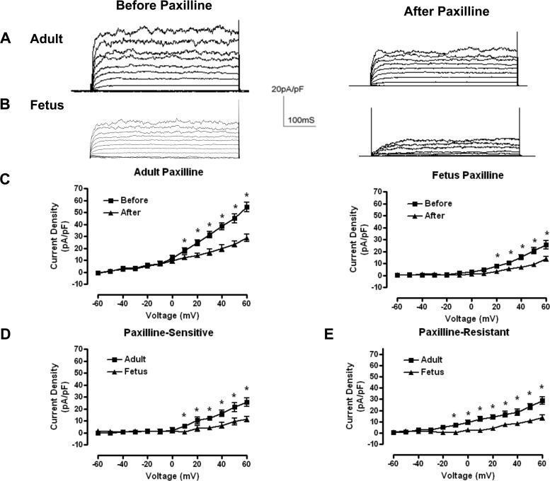
Whole-cell currents from long-term hypoxia (LTH) adult and fetal smooth muscle cells. A and B: representative whole-cell outward membrane current density traces elicited by a series of 10-mV depolarizing steps (−60 to +60 mV) from a holding potential of −60 mV. Traces before (left) and after (right) paxilline application are shown in typical isolated LTH adult (A) and fetal (B) basilar artery myocytes. Whole-cell current density is obtained from normalized whole-cell currents to membrane capacitance to account for size differences between adult and fetal myocytes. C: averaged steady-state current-voltage plot of outward current density in myocytes obtained from LTH adult (n = 6) and fetal (n = 7) basilar arteries before and after treatment with 5 × 10⁻⁷ M paxilline. D and E: averaged steady-state paxilline-sensitive large-conductance Ca2+-activated K+ channel (BK) currents (left) and residual, paxilline-insensitive currents (right) obtained from digital subtraction of the individual traces such as in A and B. *Significant difference with P < 0.05.

