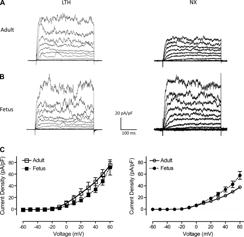
Fig. 10.
Perforated-patch, whole-cell outward current density recordings. A and B: representative whole-cell outward membrane current density traces are shown from isolated LTH and normoxic (NX) adult (A) and fetal (B) basilar artery myocytes. Currents were elicited by a series of 10-mV depolarizing steps (−60 to +60 mV) from a holding potential of −60 mV. Whole-cell current density was used to normalize whole cell currents for size differences between adult and fetal myocytes. C: averaged steady-state current-voltage plot of outward current density in myocytes obtained from LTH (left) adult (n = 5) and fetal (n = 6) and normoxic (right; taken from Ref. 3) adult (n = 4) and fetal (n = 5) basilar arteries.

