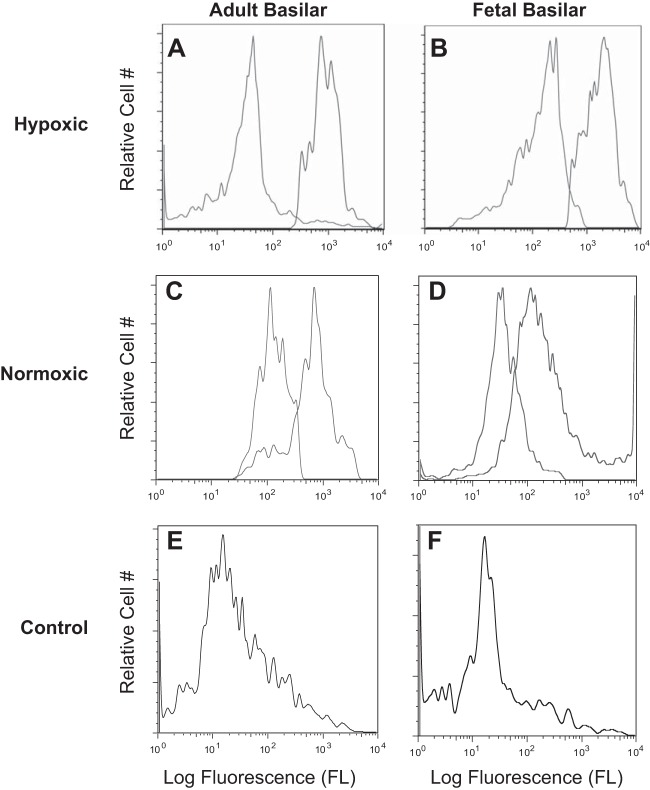
Fig. 5.
Representative flow cytometric distributions of cell surface BK channel βl subunit. A–D: isolated, intact basilar artery smooth myocytes were treated with either primary anti-BK βl (black trace) plus secondary antibody or with secondary antibody alone (gray trace). E and F: primary anti-BK βl antibody was pre-incubated with 70-fold molar excess βl epitopic peptide overnight on ice. Isolated, intact basilar artery smooth myocytes then were treated with the primary antibody and peptide mixture followed by secondary antibody to serve as antibody specificity controls (gray trace). A: LTH adult (n = 8); B: LTH near-term fetus (n = 9); C: normoxic adult (n = 13); D: normoxic near-term fetus (n = 13); E: normoxic adult (n = 13); and F: normoxic fetus (n = 13).

