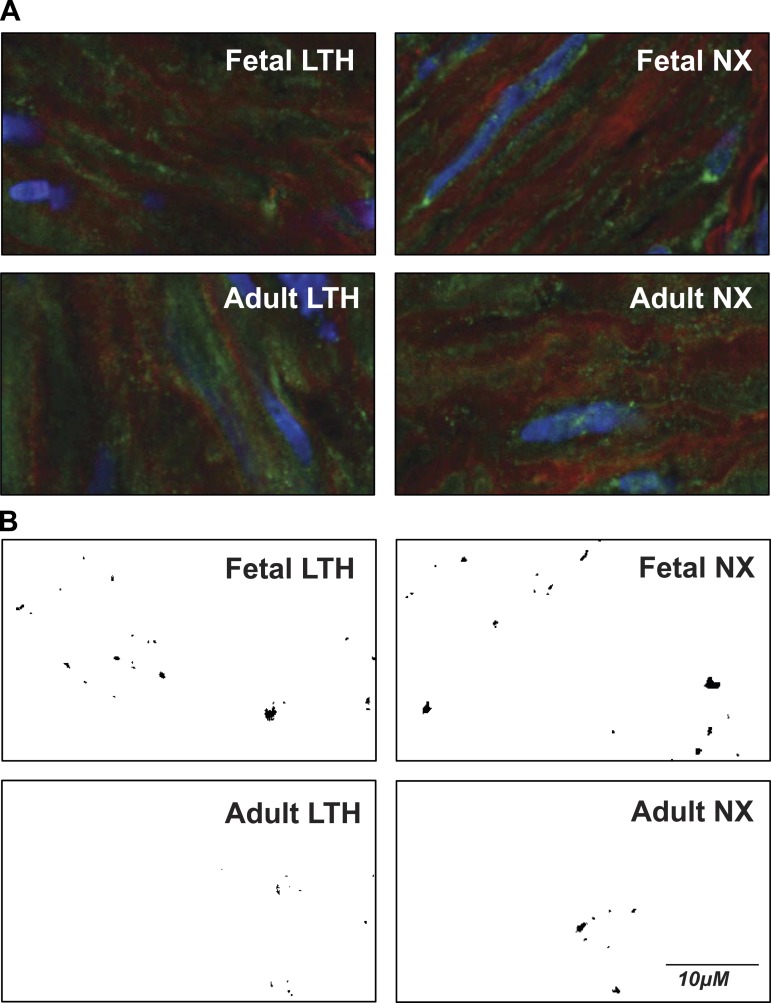
Fig. 7.
Representative confocal microscopic images of arterial myocytes reveal presence of dispersed and clustered BK channels. A: representative color images from adult LTH, fetal LTH, adult NX (normoxic), and fetal NX. Viewed areas measure 20 × 40 μm. Green color indicates presence of BK channels. B: green channel (BK fluorescence) intensities converted to binary image from same areas as above (A) after masking out all values below threshold (3.5× mean intensity). BK clusters show as black areas of different size and shape. Controls with secondary antibody alone or with primary antibody pre-absorbed with antigenic peptide revealed little to no detectable BKα fluorescence (data not shown).

