Fig. 8.
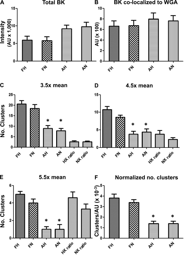
Total BK channel density, BK surface density, and BK clustering measured in confocal images of intact basilar artery myocytes. A: total BK fluorescence intensity in arbitrary units (AU; means ± SE; n = 5), where FH is fetal hypoxic (LTH), FN is fetal normoxic, AH is adult hypoxic, and AN is adult normoxic. B: BK colocalized with the surrogate surface membrane marker, wheat germ agglutinin (WGA; n = 6). C: number (No.) of BK clusters measured at 3.5 times above mean intensity (n = 7). D: number of BK clusters measured at 4.5 times above mean intensity (n = 6). E: number of BK clusters measured at 5.5 times above mean intensity (n = 6). F: number of BK clusters at 3.5 times mean intensity per total BK intensity (n = 6). Imaged areas measured 20 × 40 μm. Number of animals in each group was either 3 or 4. *Significant difference with P < 0.001 relative to either fetal group. HX ratio and NX ratio in C, D, and E refer to FH:AH and FN:AN, respectively.

