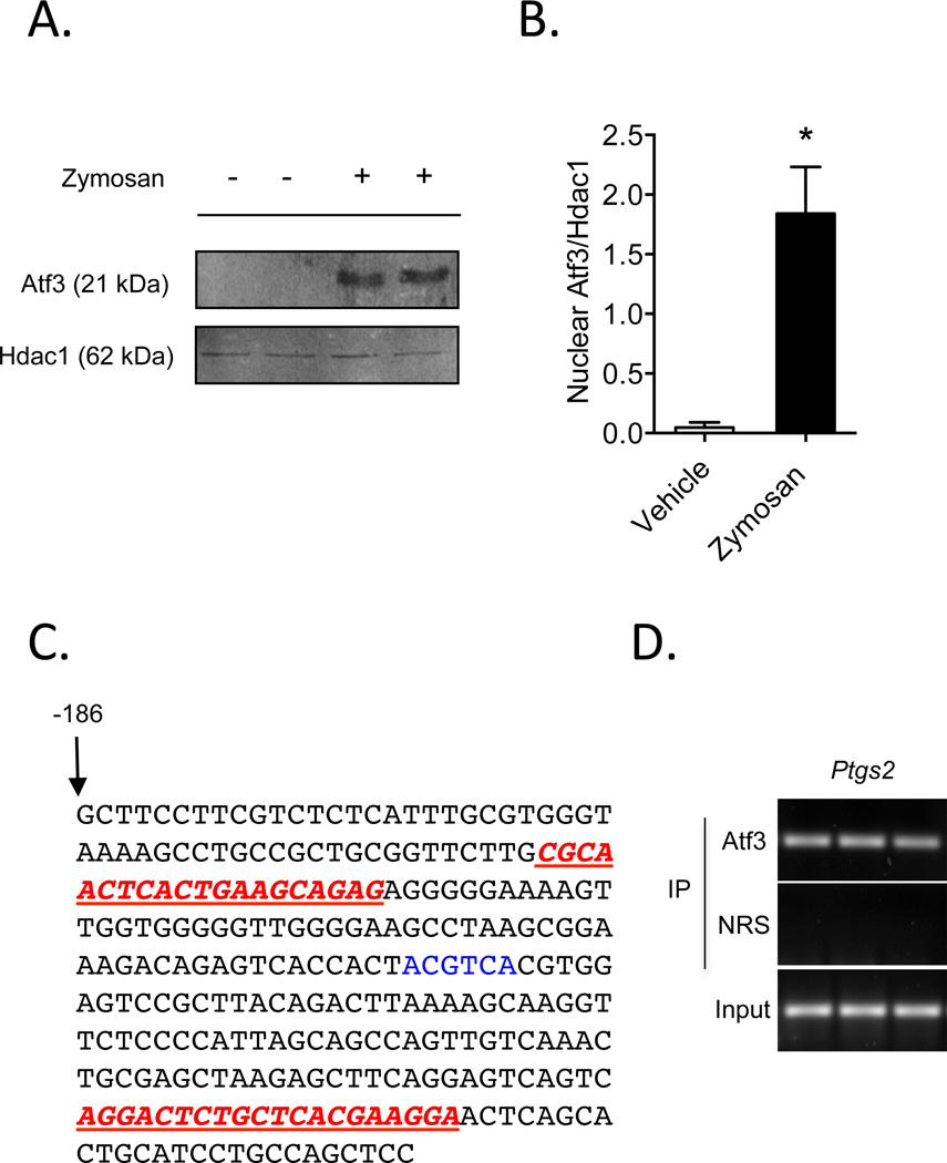
Figure 2. Atf3 is recruited to the murine Ptgs2 promoter in activated macrophages.
(A) Nuclear localization of Atf3 in zymosan-stimulated RAW 264.7 macrophages. Histone deacetylase (Hdac)-1 is shown as a control for nuclear isolation and quantification of band intensities is shown in panel B. (C) The abbreviated sequence of the Ptgs2 promoter, with primers used for the ChIP assay (underlined in red) and an ATF/CRE binding site (highlighted in blue) shown. (D) ChIP analysis of Atf3 bound to the Ptgs2 promoter in macrophages stimulated with zymosan, with non-immune rabbit serum (NRS) control and total DNA input (3%) shown. Results are mean ± SEM, n=3/group. *P<0.05

