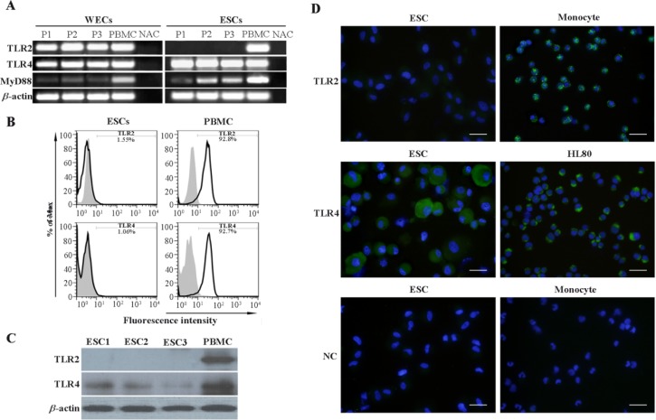
Figure 2.
Assessment of TLR2, TLR4 and MyD88 expression by ESCs and WECs. A: Assessment of TLR2, TLR4 and MyD88 transcripts expression by ESCs and WECs using RT-PCR. B: Flow cytometric analysis of TLR2 and TLR4 expression by ESCs. C: Western blot analysis of TLR2 and TLR4 expression by ESCs. In RT-PCR and Western blot analyses, PBMC was used as positive control. Monocyte gate of PBMC served as positive area in flow cytometry. D: Immunofluorescent staining of TLR2 and TLR4 in ESCs. Monocytes and HL60 cells were used as positive cell controls for TLR2 and TLR4 immunofluorescent stainings, respectively. Reagent negative control (NC) slides received isotype- matched preimmune normal serum. Nuclei were counterstained with DAPI. P1-3: Three representative participants 1-3, ESCs1-3: Endometrial stromal cells from three representative participants, PBMC: Peripheral blood mononuclear cells, NAC: No amplification control

