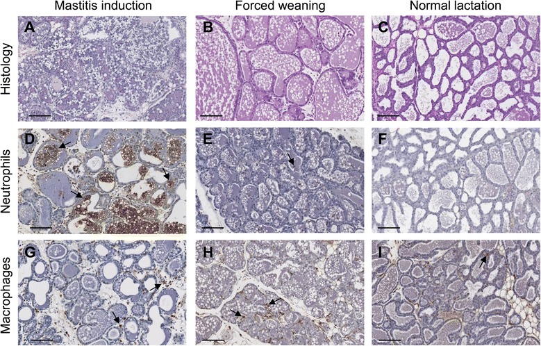
Figure 1.

Alterations in cellular components of the mouse mammary gland during mastitis induction and forced weaning. Figure shows tissue histology (A-C, haematoxylin and eosin stained sections) and abundance of neutrophils (D-F) and macrophages (G-I) 24 hours after mastitis induction or forced weaning. The macrophages and neutrophils are stained brown, and counter-stained with haematoxylin and indicated by arrows. If the mastitis-inducing agent is administered at the same time as forced weaning, it can be difficult to distinguish the specific inflammatory response to mastitis from the inflammatory response to forced weaning, as both cause alterations in immune cells. Magnification x20, scale bars represent 100 μm. Adapted from Glynn et al. [47] with permission.
