Figure 2.
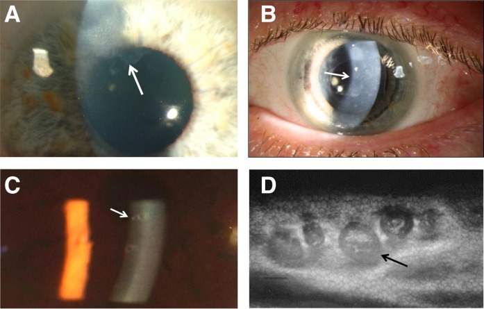
Corneal abnormalities. (A) Mild scarring caused by recurrent corneal erosions shown on slit-lamp examination in a man with X-linked Alport syndrome (arrow), renal failure, and perimacular retinopathy. The patient’s mother is also affected with renal disease and similar corneal changes. (B) Posterior polymorphous corneal dystrophy (arrow) with diffuse and vesicular lesions posteriorly at the level of Descemet’s membrane on slit-lamp examination in a man with X-linked Alport syndrome, renal failure, lens replacement for lenticonus, and perimacular retinopathy. (C) A slit-lamp view of posterior polymorphous corneal dystrophy showing the characteristic doughnut-like vesicles posteriorly (arrow). (D) Specular microscopy of the corneal endothelium in the patient in C showing that the doughnut-like lesions are vesicles with thick dark borders around clusters of endothelial cells (arrow).

