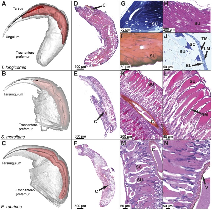
Fig. 2.
Comparative morphology of the venom apparatus of T. longicornis (Top), S. morsitans (Middle), and E. rubripes (Bottom). (A–C) Three-dimensional MRI reconstruction showing forcipule (white), venom gland (red), and chitinous duct and calyx. (D–F) Low-magnification overview of transverse sections of venom glands stained with H&E (D and F) or PAS (E) showing differences in the arrangement of secretory units in relation to the calyx. (G–J) High-magnification image showing organization of secretory units in the venom gland of T. longicornis: (G) Toluidine blue-stained diagonal section of intact forcipule showing attachment of the calyx to the cuticle (asterisk) and tightly packed secretory units (SU) connected to the lumen through pores in the calyx (C). (H) Jones-PAS stained transverse section displaying secretory units lined with darkly stained nuclei of intermediary cells. (I) Eosin-stained section showing point of termination at the basal lamina and wide diameter of each secretory unit. (J) Alcian yellow-stained section of same region with darkly stained secretory cells (SC), basal lamina (BL), transverse muscle (TM), and longitudinal muscle (LM) layers. (K–N) Magnified sections of E and F showing details of venom gland organization in S. morsitans and E. rubripes along with (L) the presence of radial muscle fibers (RM) and (N) the valve-like structure (V) connecting the secretory unit to the lumen.

