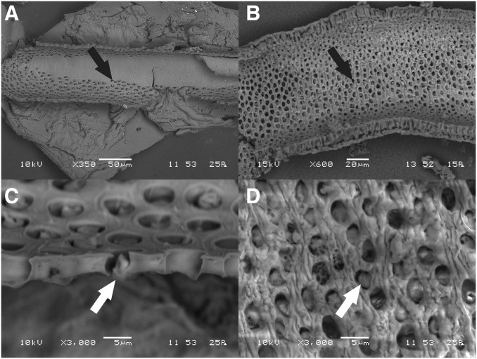
Fig. 3.
Inner structure of the calyx. SEM micrographs of the inner wall of the calyx of (A and C) T. longicornis and (B and D) S. morsitans, showing the density of pores (black arrow) and hence number of individual secretory units. Secretory units open into the lumen through these pores by a distal canal cell forming a one-way valve (white arrow), remnants of which can be seen in each of the high-magnification images (C and D). The transition from the nonporous duct to the porous calyx can be observed in A.

