Fig. 4.
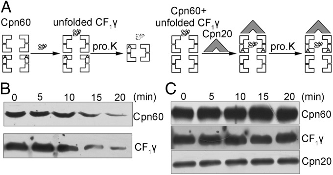
Proteinase K digestion analysis of Cpn60/Cpn20-assisted refolding of CF1γ. (A) Schematic representation of molecular interactions during the experiment. The Cpn60 and unfolded CF1γ were incubated in the absence (Left) or presence (Right) of Cpn20 and ADP for 10 min. Following these steps, the protein mixtures were treated with proteinase K for 0–20 min. (B) Immunoblot analysis of CF1γ after proteinase K treatments corresponding to the procedure described in A, Left. The proteins were separated by 15% (wt/vol) SDS/PAGE, and the Cpn60 degradation product can be detected on 7% (wt/vol) SDS/PAGE. (C) Immunoblot analysis of digestion of CF1γ by proteinase K corresponding to the procedure described in A, Right. The experimental analysis was the same as in B.

