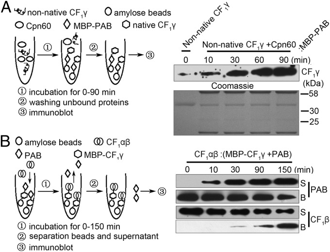
Fig. 5.
Functional analysis of PAB in the assembly of CF1γ into the ATP synthase CF1 catalytic core. (A) Pull-down assay of the interaction between PAB and unfolded or Cpn60-assisted refolded CF1γ. The experimental procedure is diagrammed (Left). The unfolded CF1γ was incubated with Cpn60/Cpn20 for 0, 10, 30, 60, and 90 min in the presence of ATP and then subjected to pull-down assay with PAB. The Coomassie-stained gel is shown below the immunoblot (Right, Lower). (B) Analysis of substitution during CF1γ assembly with CF1αβ. PAB and CF1γ-MBP formed a PAB–CF1γ complex bound to amylose beads via the MBP tag on CF1γ, and then the CF1αβ complex was added, followed by incubation for 0, 10, 30, 90, and 150 min. Proteins in the supernatant (S) and bound to the amylose beads (B) were recovered and subjected to immunoblot analysis.

