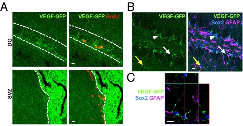
Fig. 1.
Adult hippocampal NSPCs express VEGF in vivo. (A, Top) Images from adult VEGF-GFP mice showing GFP+ puncta lining the SGZ and surrounding cells labeled with BrdU 2 h before euthanasia. (A, Bottom) SVZ showed weaker GFP expression than the SGZ, whereas the CP showed intense GFP expression. (B) GFP+ puncta were found surrounding Sox2+ TAPs (arrowheads) and filling Sox2+/GFAP+ RGLs (white arrow) in the SGZ. GFAP+ cells with astrocytic morphology also colabeled with GFP (yellow arrow). (C) Orthogonal image of a single 1-μm z-slice showing GFP+ puncta colocalizing with GFAP in a GFAP+/Sox2+ RGL. (Scale bars: 10 μm.)

