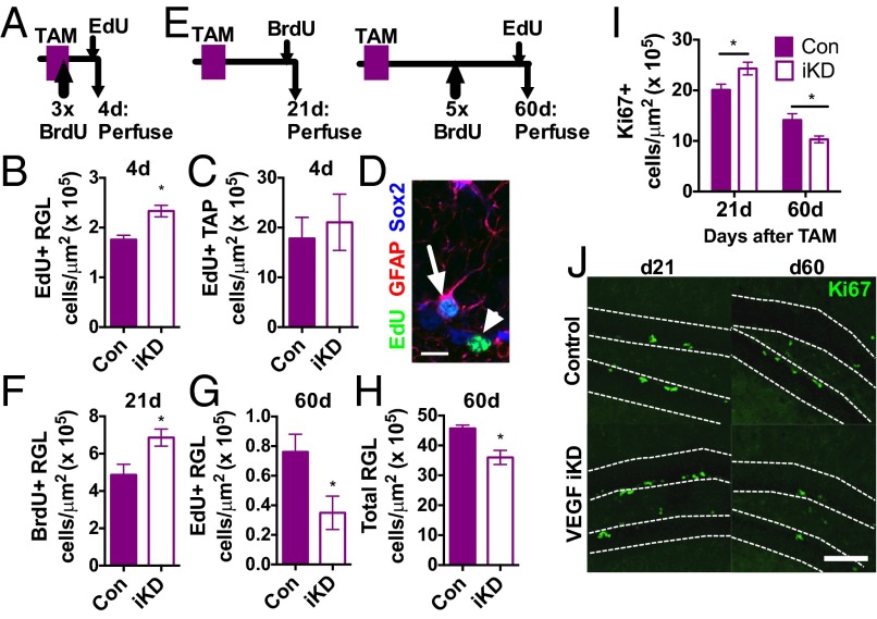
Fig. 4.
Loss of NSPC-derived VEGF disrupts NSPC self-regulation in vivo. (A) Adult control (n = 3) and VEGF-iKD mice (n = 7) were treated with TAM for 5 d and then perfused 4 d later. (B) VEGF-iKD led to an increase in the number of EdU+ proliferating RGL stem cells. *P = 0.017, Mann–Whitney test. (C) VEGF-iKD did not significantly alter the number of proliferating TAPs. P > 0.1, Mann–Whitney test. (D) Example EdU+/GFAP+/Sox2+ RGLs (arrow) and EdU+/Sox2+ TAPs (arrowhead) in a 1-μm z-slice. (Scale bar: 10 μm.) (E) Adult control and VEGF-iKD mice were treated with TAM for 5 d and then perfused after 21 d (n = 10 control and n = 11 VEGF-iKD mice) or 60 d (n = 4 control and n = 10 VEGF-iKD mice). (F) VEGF-iKD increased proliferation of the RGL stem cell population 21 d after knockdown. *P = 0.0124, t test. (G) Sixty days after knockdown, RGL stem cell proliferation was decreased in VEGF-iKD mice relative to controls. *P = 0.0465, Mann–Whitney test. (H) At 60 d, the number of GFAP+/Sox2+ RGLs was decreased in VEGF-iKD mice relative to controls. *P = 0.024, Mann–Whitney test. (I) VEGF-iKD led to an increase in Ki67+ proliferating cells in the SGZ after 21 d (d21) but a decrease after 60 d (d60) (two-way ANOVA: interaction, P = 0.0042; day, P < 0.0001; genotype, P = 0.94). Post hoc planned comparisons within day (21 d: P = 0.0343, t test; 60 d, P = 0.0140, Mann–Whitney test). (J) Example images of proliferating Ki67 cells in control and VEGF-iKD mice at day 21 and day 60. (Scale bar: 100 μm.) Data represent mean ± SEM.

