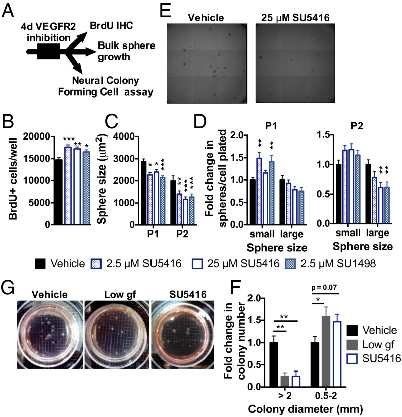
Fig. 6.
Inhibition of VEGFR2 signaling disrupts NSPC self-regulation. (A) NSPCs were treated in a monolayer for 4 d with VEGFR2 kinase inhibitors: SU5416 or SU1498. (B) Number of BrdU+ proliferating NSPCs was increased by exposure to either SU5416 or SU1498 (ANOVA: P = 0.0010; n = 3–5 wells per group per experiment; three experiments). (C) Four days of VEGFR2 inhibition led to a smaller sphere size over subsequent passages (two-way ANOVA: interaction, P = 0.46; passage, P < 0.0001; VEGFR2 inhibition, P < 0.0001; n = 6 wells per group per experiment; three experiments). (D) Formation of small spheres (diameter ≤100 μm) was enhanced in P1 after VEGFR2 inhibition. In P2, formation of large spheres (diameter >100 μm) was inhibited (P1, two-way ANOVA: interaction, P = 0.0029; size, P < 0.0001; VEGFR2 inhibition, P = 0.24; P2, two-way ANOVA: interaction, P = 0.0012; size, P < 0.0001; VEGFR2 inhibition, P = 0.31). (E) Example images of spheres after P2. (Grid: 1.5 μm.) (F) Neural colony-forming cell (NCFC) assay revealed that treatment with 5 ng/mL EGF/FGF2 [low growth factor (gf)] or 25 μM SU5416 decreased formation of stem cell-derived colonies with diameter >2 mm and caused a trend toward an increase in smaller, progenitor-derived colonies (two-way ANOVA: interaction, P = 0.0019; size, P < 0.0001; treatment, P = 0.90; n = 2–3 wells per experiment; three experiments). All post hoc comparisons were Dunnett’s comparisons to vehicle: *P < 0.05; **P < 0.01; ***P < 0.001. Data are mean ± SEM normalized to vehicle within experiment. (G) Example NCFC spheres after 3 wk in culture. (Grid: 2 mm.)

