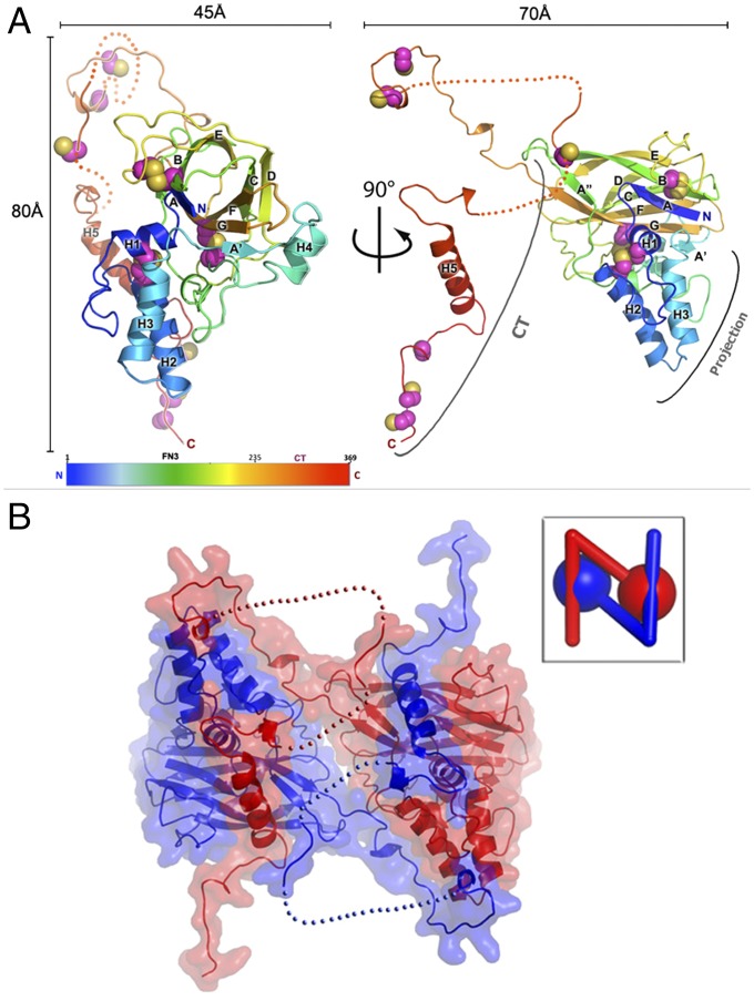
Fig. 1.
The structure of fusolin. (A) The structure of fusolin is represented as a ribbon diagram colored in a blue–red gradient from the N to C termini. Cysteine residues are shown as spheres. (B) Fusolin forms a domain-swapped dimer shown as a ribbon diagram within a semitransparent molecular surface. (Inset) A schematic representation of the dimer.

