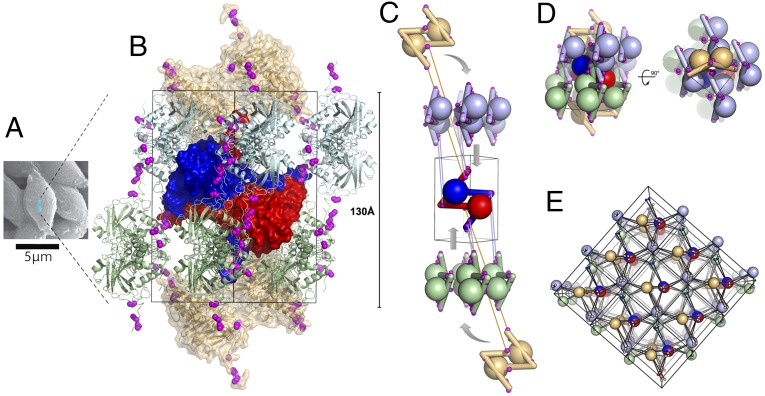
Fig. 3.
Spindles are single-chain crystalline polymers of fusolin dimers crosslinked by a 3D network of disulfide bonds. (A) Scanning electron micrograph of spindles purified from EV-infected cockchafer larvae. The blue box represents a unit cell (not to scale). (B) Representation of a spindle unit cell viewed along a twofold crystallographic axis. Cysteines involved in interdimer crosslinks are shown as magenta spheres. The central dimer is represented as a blue-red molecular surface; two crowns of four dimers are shown in green and cyan (one dimer of each crown is masked because of its location on the opposite side of the unit cell). The two capping dimers are shown in brown as a semitransparent molecular surface. (C) Schematic representation of the unit cell assembly. Interdimer disulfide bonds involving the central dimer are indicated by links between the interacting cysteines. (D) The full unit cell is shown in the same orientation as in C and in an orthogonal view. (E) Representation of 18 unit cells with each dimer shown as a large sphere and cysteines as smaller spheres. Intersphere links highlight the 3D network of covalent bonds crosslinking spindles.

