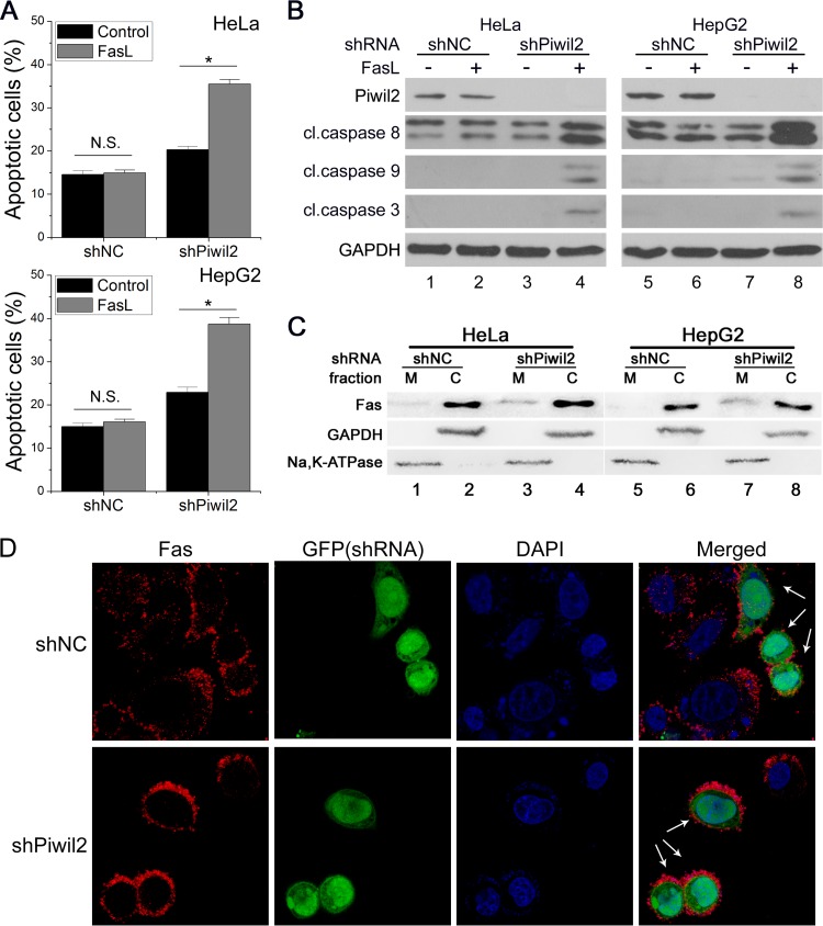
FIG 1.
Piwil2 loss sensitizes cells to Fas-mediated apoptosis. (A and B) NC (shNC) and Piwil2 knockdown (shPiwil2) cells were mock treated or treated with FasL (100 ng/ml) and then harvested for apoptosis analysis using FACS assay and Western blot analysis. Error bars indicate SE (N.S. [not significant], P > 0.05; *, P < 0.05). cl., cleaved. (C and D) The impact of Piwil2 depletion on the membrane localization of Fas was determined by membrane (M)/cytosolic (C) fractionation (C) and immunofluorescence staining (D). Na, K ATPase (membrane) and GAPDH (cytosolic) were used as controls. Green fluorescent protein (GFP) is expressed from shRNA vectors and indicates the cell outline. The arrows indicate the expression of the shRNA vectors. Anti-Fas was used for panel D.

