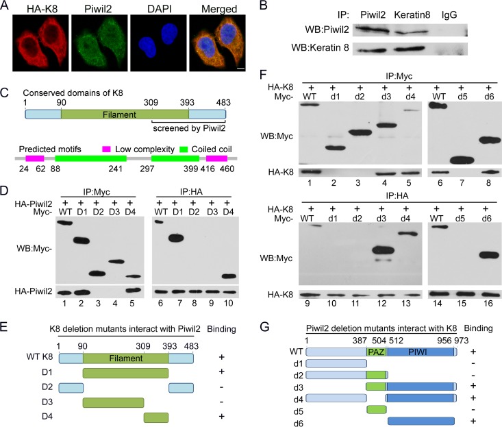
FIG 2.
Interaction of Piwil2 and K8. (A) Piwil2 and K8 are colocalized mostly in cytoplasm. Cells were transfected with HA-K8 vector and harvested for immunofluorescence assay with anti-Piwil2 and mouse anti-HA antibodies. Scale bar, 5 μm. (B) Endogenous interactions between Piwil2 and K8 in HeLa cells. Coimmunoprecipitation was performed with anti-Piwil2 or anti-K8, followed by Western blotting. (C) Filament domain of K8 and the predicted domains by SMART analysis. (D) Interaction between HA-tagged Piwil2 and different Myc-tagged K8 mutants. (E) Schematic of different K8 deletion mutants. (F) Interaction between HA-tagged K8 and different Myc-tagged Piwil2 mutants. (G) Schematic of different Piwil2 mutants. All of these assays were performed in HeLa cells. WB, Western blotting; WT, wild type.

