FIG 3.
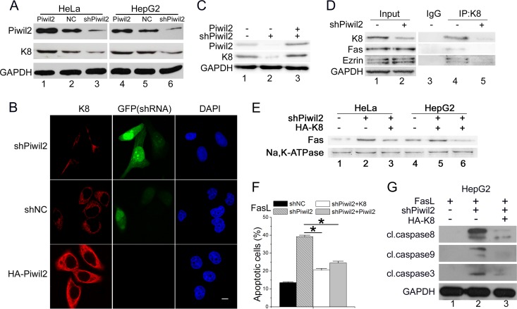
Piwil2 increases K8 protein levels. (A) HeLa and HepG2 cells were transfected with Myc-Piwil2, shNC, or shPiwil2 vector. After 48 h, cell lysates were used for Western blotting with anti-K8. (B) Piwil2 knockdown decreased the K8 fluorescence intensity significantly. HeLa cells were transfected with shNC or shPiwil2 vector and harvested for immunofluorescence assay with anti-K8 antibodies. GFP is expressed from shRNA vectors. Scale bar, 5 μm. (C) Rescue experiments of the knockdown of Piwil2 on the protein level of K8. HepG2 cells were transfected with shPiwil2 or Piwil2 expression vector as indicated. (D) Piwil2 knockdown decreased the interaction between K8 and Fas-ezrin complex. HeLa cells were transfected with shNC or shPiwil2 vector, and a coimmunoprecipitation assay was performed using anti-K8, followed by Western blotting. (E) The impact of K8 on Fas localization in Piwil2 knockdown cells. The level of membrane Fas was determined by membrane protein separation followed by Western blotting. (F) Both Piwil2 and K8 overexpression reduced the sensitivities of Fas-mediated apoptosis enhanced by Piwil2 knockdown. (G) Western blot analysis of caspases activation after FasL induction. Cells were transfected with shNC, shPiwil2, pcDNA3.1, HA-K8, and Myc-Piwil2 plasmids as indicated 48 h before Western blotting. Cells were treated with FasL (100 ng/ml) as indicated. Error bars indicate SE (*, P < 0.05).

