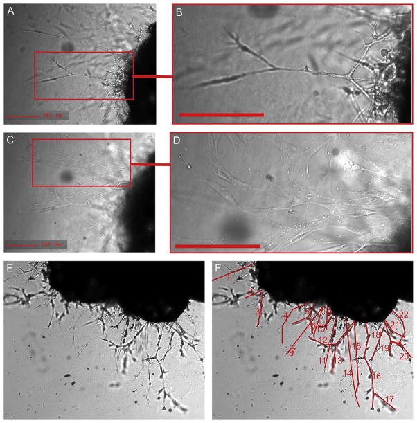
Figure 5.2.
Cells emerging from mouse adipose tissue explant. (A, B) Capillary sprout emerging from embedded mouse explant, displaying characteristic linear branching structure. (C, D) Focus set to the surface of the well, where fibroblastic adherent cells can be seen emerging from the explant, observed at a different optical plane of the image. (E) Phase contrast image of the explant and the capillary sprouts 14 days post-embedding. (F) Structures shown in red highlight formations that can be considered to be sprouts.

