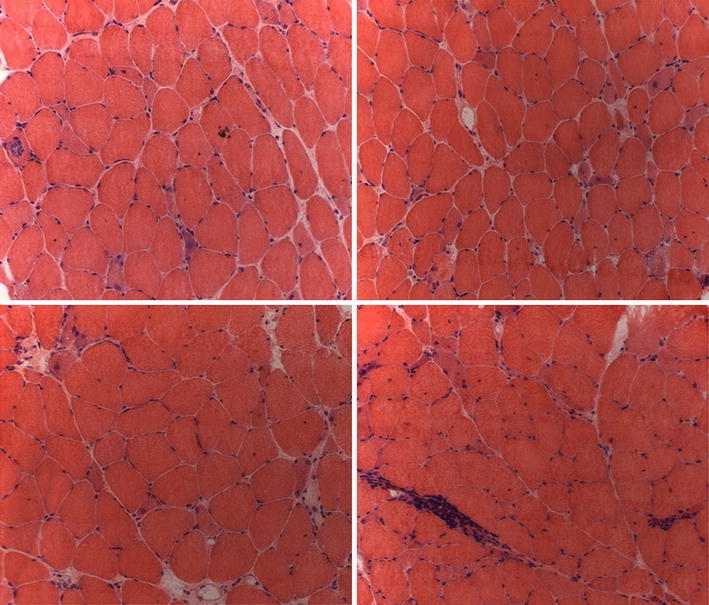
Fig. 1.

Muscle biopsy from a patient with polymyositis and anti-HMGCR autoantibodies (hematoxylin–eosin stain, original magnification ×20). The biopsy shows scattered necrotic fibers, some of which invaded by mononuclear cells, basophilic regenerating fibers and an inflammatory infiltrate in the perimysium. (Courtesy of Dr. Vattemi G.)
