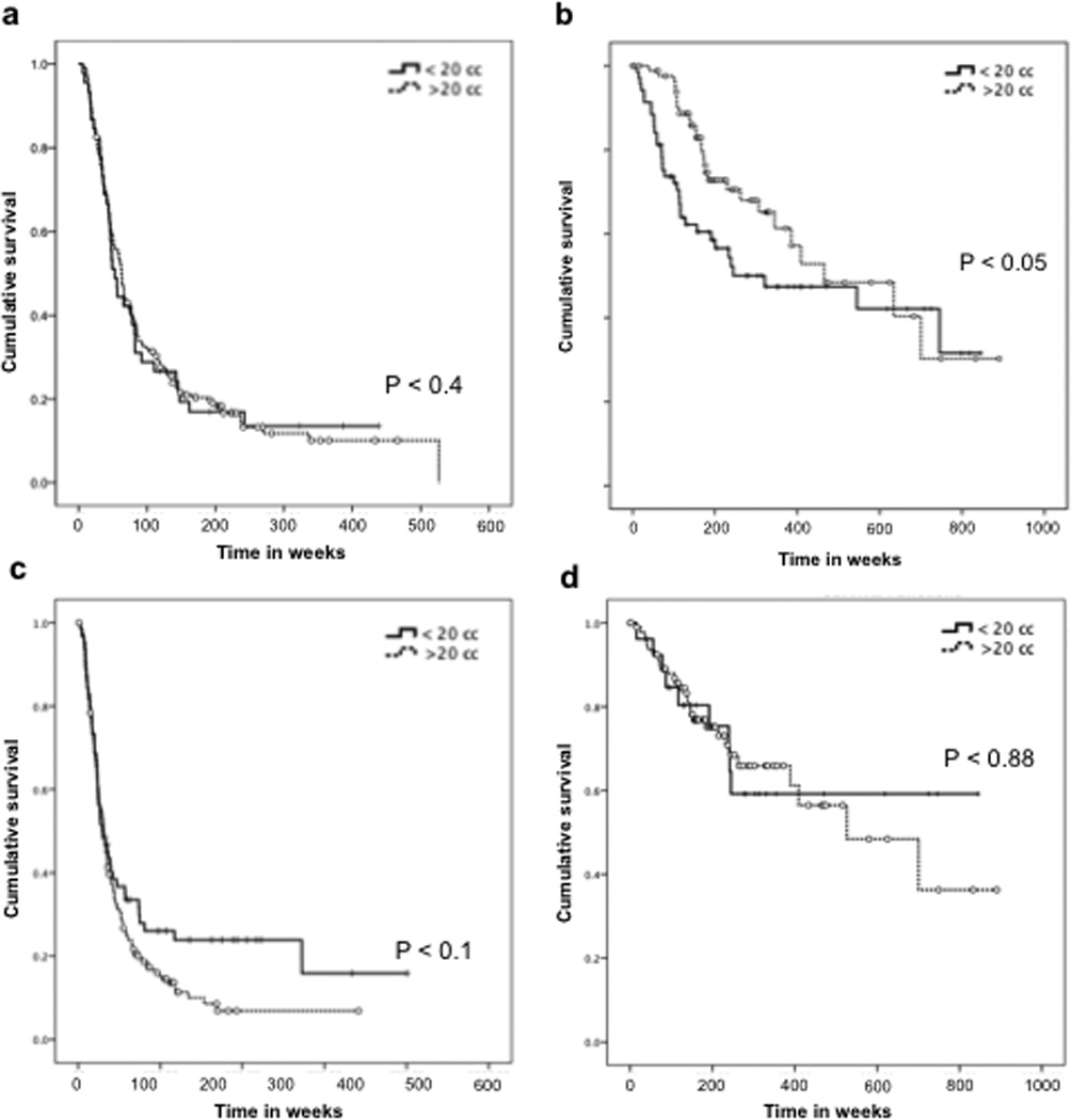
Fig. 2. Survival analysis of malignant astrocytomas based on histopathology and IDH1 status.

Kaplan-Meier curves show that in patients with histological diagnosis of GBM (A), the overall survival is similar with resection volumes > 20 cc or < 20 cc. However, in patients diagnosed with AA (B), resection volume < 20 cc correlated with worse prognosis (P < 0.05). Neither IDH1 WT (C) nor IDH1 mutants (D) demonstrated significant differences in patient survival between the two resection volume groups.
