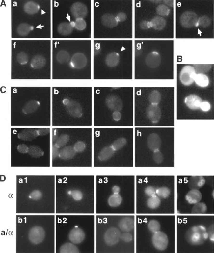
Fig. 1.
Localization of Bud5-GFP in haploid and diploid cells. (A) Localization of Bud5-GFP in haploid α cells. Approximately 150 cells were examined for each panel in Figs. 1 and 2, except where noted. Representative micrographs are shown. Bud5-GFP images in yeast cells (strain HPY307) at different stages of the cell cycle are shown (panels a through e). Panels f through g’ show two pairs of images of Bud5-GFP (f and g) and Calcofluor staining (f’ and g’) of unbudded cells. The arrowheads indicate the Bud5-GFP signal in a patch at the incipient bud site in G1 cells; the arrows indicate the Bud5-GFP signal in a small bud. (B) Overexpression of Bud5-GFP in haploid a cells. Bud5-GFP images of a bud5Δ (strain IH2423) cells expressing BUD5-GFP from a multicopy plasmid are shown. (C) Localization of Bud5-GFP in diploid a/α cells (strain HPY309). A small percentage (5%) of cells with small-sized buds also showed the Bud5-GFP signal at the opposite pole of the mother cell (panel b), in addition to buds. (D) Localization of Bud5-b1-GFP in haploid a and diploid a/α cells. Approximately 200 cells were examined for each panel. Strain HPY378 was used in panels a1 through a5, and strain HPY388 was used in panels b1 through b5. Calcofluor staining was shown in panels a5 and b5. Bud5-b1-GFP sometimes localized in a patch at the neck in a/α cells (panel b4), which was not observed in wild-type cells.

