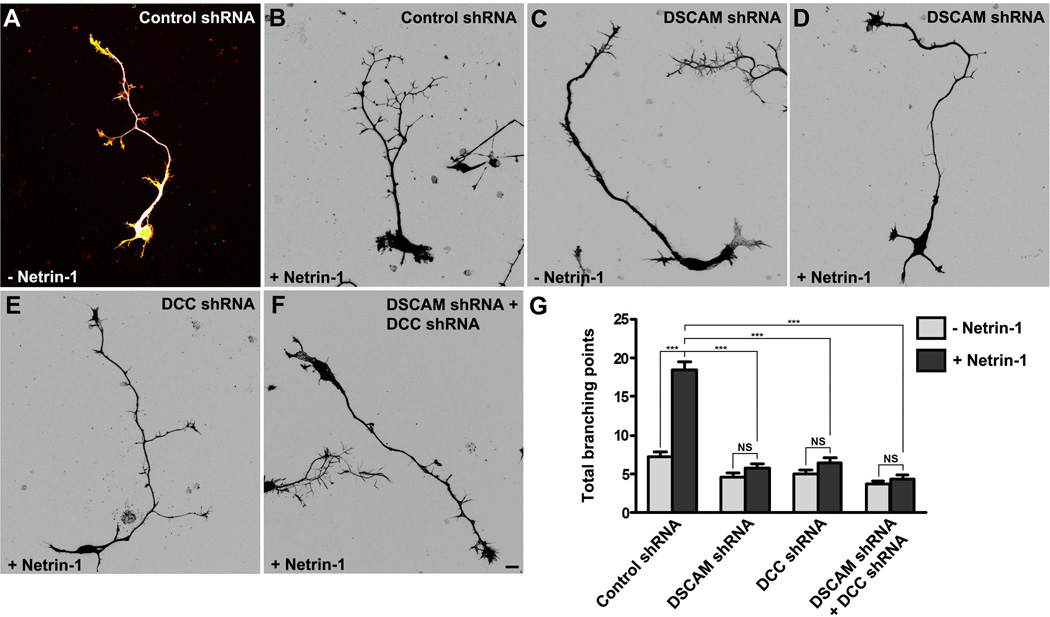
Figure 6.
DSCAM collaborates with DCC in Netrin-1-induced axon branching. (A–F) E15 mouse cortical neurons were transfected with Venus YFP plus control shRNA (A–B), Venus YFP plus DSCAM shRNA (C–D), Venus YFP plus DCC shRNA (E), and Venus YFP plus DC shRNA and DSCAM shRNA (F), respectively. Primary neurons were cultured in the presence (B, D, E–F) or absence (A and C) of purified Netrin-1 for 4 d and stained with anti-tau, phalloidin (F-actin) and DAPI. Images (B–F) are inverted LUT (look-up table) from original RGB pictures. Scale bar, 10 µm. (G) Quantification of Netrin-1-induced axon branching. The branching point with a branch longer than 10µm was selected. Only axon branching points of YFP-positive neurons not in contact with other cells were measured and used in the statistical analyses. More than 80 neurons from three separate experiments were calculated. Data are mean ± s.e.m. ***, p<0.001 (One-way ANOVA with Kruskal–Wallis test for post-hoc comparisons). NS, not significant.

