
Fig. 5.
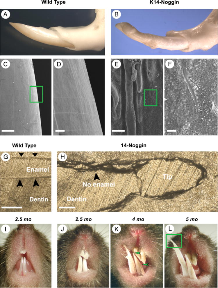
Growth abnormalities of the K14-Noggin incisors caused by the loss of enamel. (A, B) Comparative gross morphology of the mandibular incisors in the adult WT (A) and K14-Noggin mice (B). K14-Noggin incisors are thick, wide, blunt ended, and misaligned. (C–F) On SEM, the surface of the K14-Noggin incisors is rough and defective (E, F). It shows both macroscopic signs of deterioration in the form of deep, parallel ridges (E) and microscopic irregularities in the form of multiple bud-like formations (F). The surface of WT incisors is smooth (C, D). (G, H) On ground sections, WT incisors display a clear, thick layer of enamel (G). In contrast, K14-Noggin incisors do not have any enamel layer present (H). (I–L) Progressive changes of the incisors in K14-Noggin mice. Unlike WT incisors (I), K14-Noggin incisors are a dull white and deteriorate because of constant rubbing against each other (J–L). These changes start early in life and with age become more severe. The bottom incisors grew very long and became needle sharp (L). Section plane: sagittal (G, H). Scale bars: 100 μm (C, E, G, H); 20 μm (D, F).