
Figure 1. Distribution of TUNEL+ cells in developing feather buds and follicles.
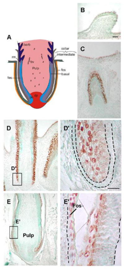
(A) Schematic representation of longitudinal feather follicle sections. There are three epidermis layers in the feather filament: the basal layer, intermediate layer, and feather sheath. The feather filament surrounds the pulp and shows continuity with the invaginated epidermis that has become the feather sheath. (B) E 9 feather buds. (C) A section of E12 invaginating feather follicles. TUNEL+ cells in regions undergo active tissue remodeling. (D) At E15 a longitudinal section of a wing feather, TUNEL+ cells are detected in the feather sheath, higher magnification of the box area is shown in (D′). Strong TUNEL+ cells are detected in the feather sheath and dermal sheath, but the basal layer, demarcated by a black dotted line, lacks TUNEL+ cells. (E) At E16, a space is created between the feather follicle and dermal sheath, higher magnification of the box area is shown in (E′), TUNEL+ cells distribute along the separated and keratinized feather sheath and dermal sheath. A feather now can emerge from its sheath. bv, blood vessels; dp, dermal papilla; Fes, feather sheath; Fos, follicle sheath; rm, ramogenic zone. Scale bars, 100 μm (B-E), 50 μm (D′, E′).