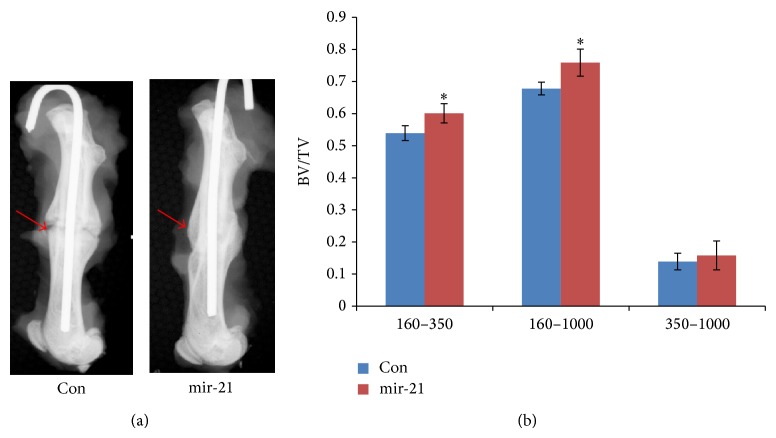
Figure 2.
mir-21-MSCs promoted fracture healing in vivo via radiologic analysis. (a) X-ray was taken 5 weeks after fracture with a high-resolution digital radiograph system (Faxitron MX-20, Illinois, USA) using an exposure of 32 kV for 10 seconds. (b) Micro-CT analysis data showed that BV/TV of newly formed bone in mir-21-MSCs group was much higher than that in control which prompted that the elevation of mir-21 accelerated the deposition of newly formed bone. Attenuation above 160 represented total mineralized tissue, and attenuation between 160 and 350 represented the newly formed calluses. * P < 0.05, compared with control.

