
Figure 3.
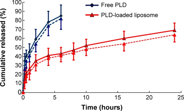
In vitro release of the polydatin-loaded liposome in phosphate-buffered saline (pH 7.4, n=3, solid line) and simulated gastric fluid (pH 1.2, n=3, dotted line).
Notes: Red line indicates polydatin (PLD) loaded liposome; blue line indicates free polydatin.