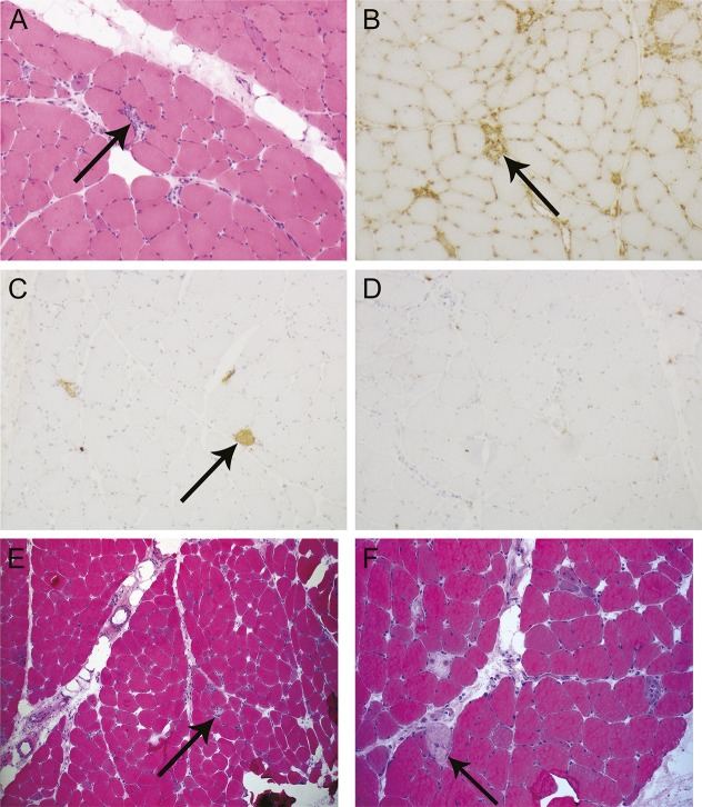
Figure 1. Muscle biopsy images demonstrating a pauci-immune necrotizing autoimmune myopathy in illustrative cases 1 and 2.
(A–D) Case 1 deltoid muscle. (A) Frozen section showing scattered necrotic fibers with associated macrophage infiltration but no discernible lymphocytic infiltration (arrow) (hematoxylin & eosin [H&E] stain, magnification 100×). (B) Immunohistochemistry for major histocompatibility complex class I highlights macrophages associated with muscle fiber necrosis and background capillaries, but there is no generalized upregulation on uninvolved muscle fibers in this biopsy sample (arrow) (magnification 100×). (C) Immunohistochemistry for complement membrane attack complex highlights foci of muscle fiber necrosis, but there is no microvascular deposition (arrow) (magnification 100×). (D) Immunohistochemistry for CD3 reveals a paucity of T cells in areas of fiber degeneration. (E, F) Case 2 deltoid muscle. (E) H&E stain (magnification 40×). (F) H&E stain (magnification 100×). There are necrotic fibers with inflammatory cells (arrow) involved in the process of myophagocytosis and a few regenerating fibers, consistent with a pauci-immune necrotizing myopathy.

