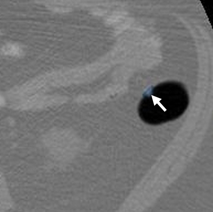
Figure 12a.

Suspected polyp. (a, b) Magnified axial 2D (a) and endoluminal 3D (b) CT colonography images show a CAD polyp candidate (arrow in a and blue area in b). The soft-tissue polypoid structure is not secondary to a normal anatomic structure or artifact and has no stool characteristics. Its maximal diameter is 6.1 mm, meeting the target lesion size threshold. This lesion may be reported as a suspected polyp and referred for optical colonoscopy for confirmation and biopsy. Alternatively, it may be conservatively managed with short-term surveillance according to the C-RADS recommendations for a patient with fewer than three 6–9-mm lesions. This lesion was reported by 11 and eight readers in the CAD-unassisted and CAD-assisted sessions, respectively.
