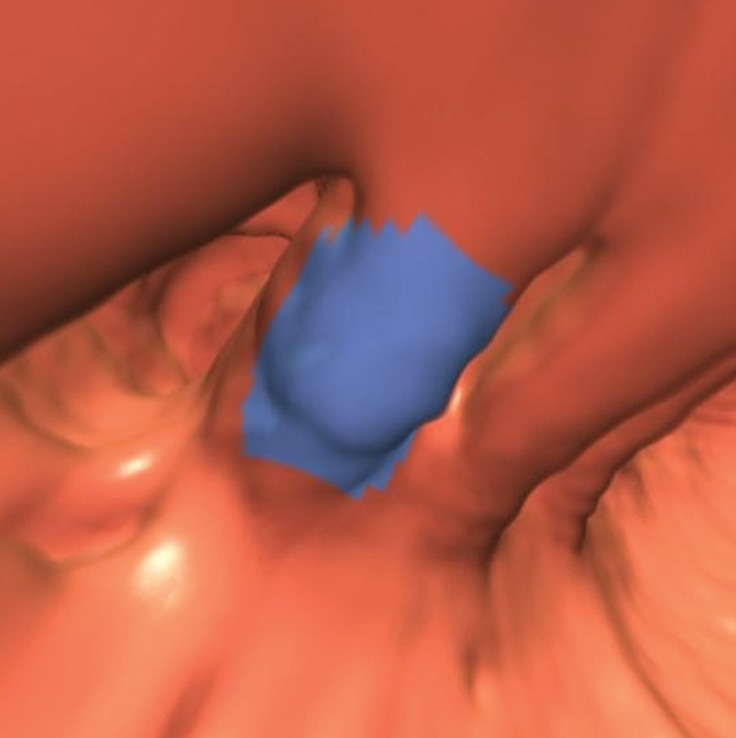
Figure 15a.

Flexural pseudotumor. Endoluminal 3D (a), magnified coronal (b), axial (c), and sagittal 2D (d) CT colonography images show a CAD polyp candidate (blue area in a and arrow in b–d) at the location of a sharp turn in the sigmoid colon. The haustral folds along the inner bend appear thickened and bulbous, a finding often referred to as a flexural pseudotumor. This example illustrates the utility of multiple projections to clearly delineate the suspicious structure. The endoluminal and axial views demonstrate that the candidate is along a sharp turn of the colon, a finding that is more difficult to appreciate on the coronal and sagittal views.
