
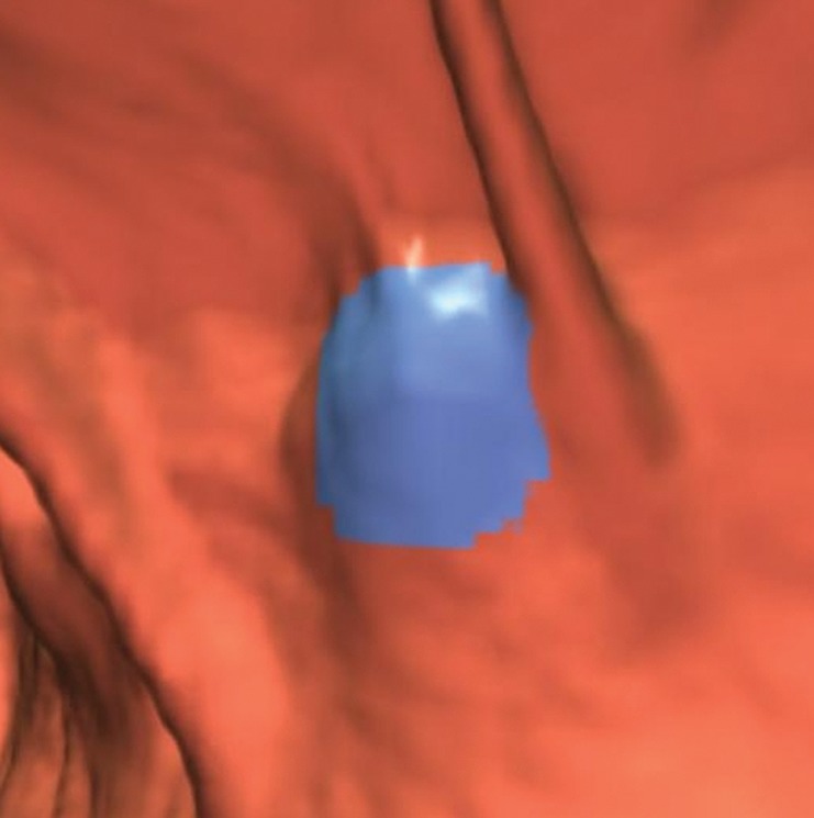
Figure 18a.
Extrinsic compression. (a, b) Endoluminal 3D (a) and magnified axial 2D (b) CT colonography images show a CAD polyp candidate (blue area in a and arrow in b) that is secondary to extrinsic compression by the anterior tip of the spleen.
Official websites use .gov
A
.gov website belongs to an official
government organization in the United States.
Secure .gov websites use HTTPS
A lock (
) or https:// means you've safely
connected to the .gov website. Share sensitive
information only on official, secure websites.
Extrinsic compression. (a, b) Endoluminal 3D (a) and magnified axial 2D (b) CT colonography images show a CAD polyp candidate (blue area in a and arrow in b) that is secondary to extrinsic compression by the anterior tip of the spleen.