
Figure 20a.
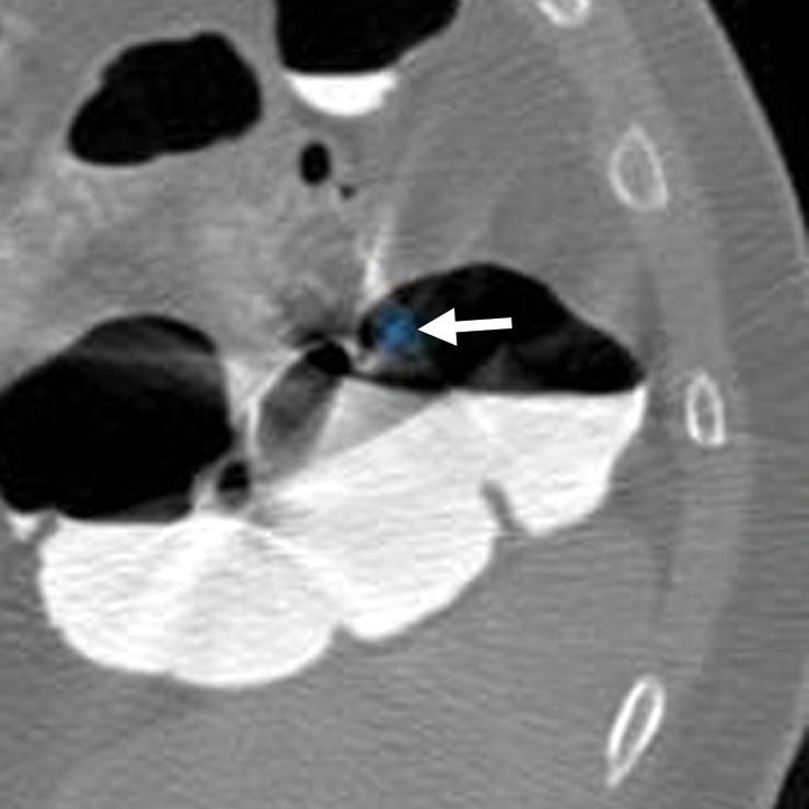
Artifacts. (a, b) Magnified axial 2D supine (a) and endoluminal 3D (b) CT colonography images show a CAD polyp candidate (arrow in a and blue area in b) that is difficult to disregard on the endoluminal view. However, the 2D view shows streak artifacts from peristalsis and the interface between air and densely-tagged liquid that may obscure large polyps. This region should be carefully evaluated on the prone view to confidently exclude polyps.