
Figure 23b.
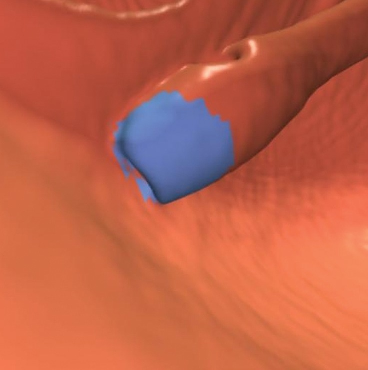
Magnified axial 2D (a) and endoluminal 3D (b) CT colonography images show a CAD polyp candidate (arrow in a and blue area in b) on the convex contour of the rectal tube. Note the symmetric ripples of the surrounding rectal mucosal surface.