
Figure 2b.
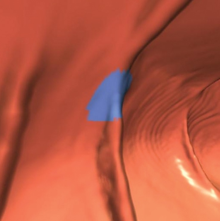
Haustral folds. (a) Endoluminal 3D CT colonography image shows a CAD polyp candidate (blue area) on a normal haustral fold. Minimal irregularity on a convex structure can generate CAD polyp candidates that are easily disregarded by the reader. (b) Endoluminal 3D CT colonography image shows a CAD polyp candidate (blue area) at the site of the convergence of two normal haustral folds that is easily disregarded by the reader.