
Figure 4a.
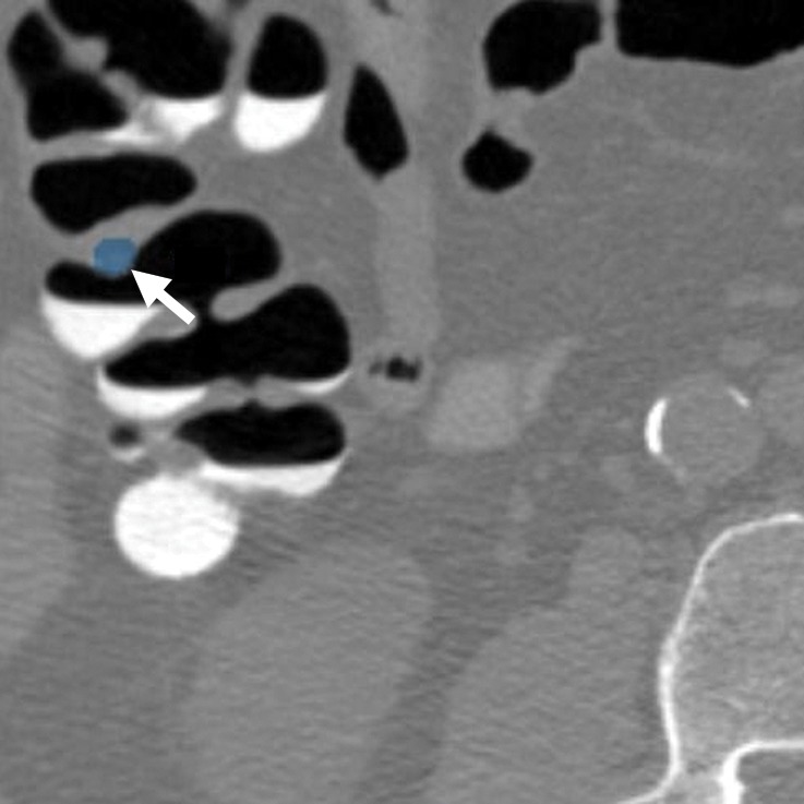
Normal anatomy. Axial 2D (a) and endoluminal 3D (b) CT colonography images show a CAD polyp candidate (arrow in a and blue area in b) at the site of a protuberance at the junction of colonic folds and the taenia coli that measures approximately 7.2 mm in its maximal dimension. This is a difficult polyp candidate to disregard, with six readers reporting it as a polyp during the CAD-assisted session and two reporting it as a polyp during the CAD-unassisted session. The location of the polyp candidate at this junction, poor distention of the colon at this site, and the bulbous appearance of adjacent folds are most consistent with normal anatomy and a CAD false-positive result.