
Figure 5d.
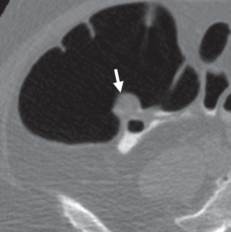
Bulbous ICV in two patients. (a) Magnified coronal 2D CT colonography image obtained in the plane of the ICV, cecum, and ascending colon shows a CAD polyp candidate (arrow) that corresponds to the lips of the ICV, which, in this patient, is sufficiently bulbous to simulate a polyp candidate. No evidence of an overlying polyp or infiltrating mass is seen. The appearance of the ICV is within the normal range and should be easily dismissed by the reader. (b) Endoluminal 3D CT colonography image shows the CAD polyp candidate (blue area) in part a. Most, but not all, of the ICV surface is marked as a CAD polyp candidate. In a primary 3D read, the colon is first viewed on an endoluminal 3D image, and the 2D image is used for problem solving. Interactive 2D and 3D viewing is needed to confirm the expected location and appearance of the ICV. On 3D images, the bulbous lips of the ICV often have polypoid morphologic characteristics and require evaluation on 2D images. (c) Endoluminal 3D CT colonography image shows a bulbous ICV that generates two CAD polyp candidates (blue area), which can be reliably dismissed with 2D problem-solving images obtained to first verify that this is the location of the ICV and, second, to ensure that there is no evidence of an overlying soft-tissue polyp or infiltrating carcinoma. (d) Axial 2D magnified CT colonography image shows the CAD polyp candidate (arrow) from part c, the terminal ileum with tagged fluid, the ICV, the cecum, and the adjacent ascending colon. The lips of the ICV are bulbous and could be concerning for a polyp in any other colonic location. However, this appearance is within the normal range for the ICV.