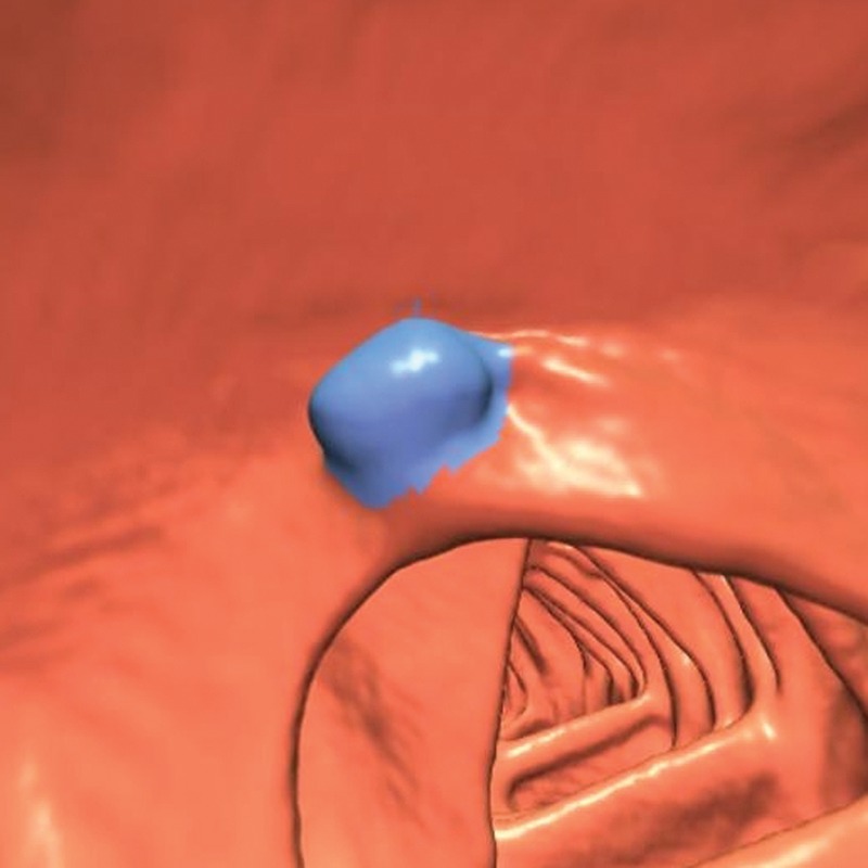
Figure 6.

Endoluminal 3D CT colonography image shows a polyp candidate (blue area) that corresponds to a markedly polypoid ICV. This polyp candidate is difficult to disregard because polyps can occur on the ICV, the appearance of which can markedly vary. Ten readers reported this finding during the CAD-assisted session, and 12 readers reported it during the CAD-unassisted session.
