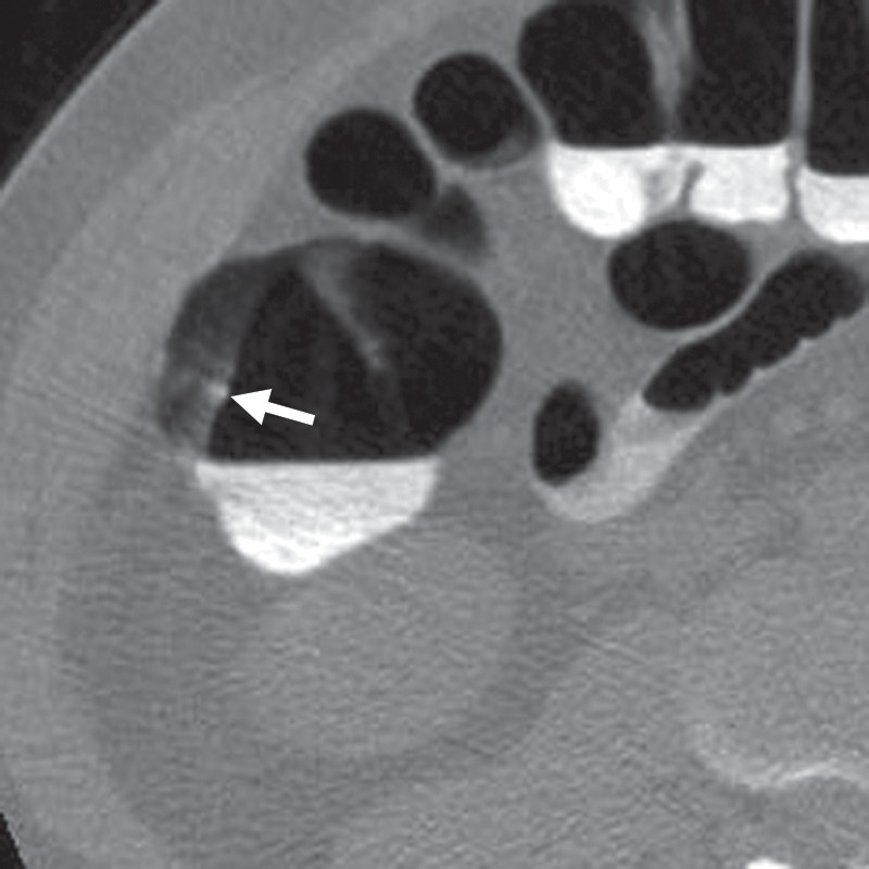
Figure 8c.

Tagged stool. (a) Endoluminal 3D CT colonography image shows a haustral fold that was marked as a CAD polyp candidate (blue area). The haustral fold has a nodular appearance due to adherent stool. Without further evaluation of the attenuation of this polyp candidate, it could be mistaken for a polyp. (b) Endoluminal 3D CT colonography image with an overlying color map shows high-attenuation (white areas) internal tagging, a finding consistent with tagged stool adhering to the normal haustral fold. (A soft-tissue polyp would be shaded red.) Care should be taken when evaluating attenuation on 2D images to confirm internal tagging rather than external surface tagging, which is sometimes seen with true polyps. (c) Magnified axial 2D CT colonography image shows the CAD polyp candidate (arrow). Interactive window width and level adjustment may be necessary to evaluate and confirm that the hyperattenuating material is internal tagging rather than the external surface tagging that is sometimes seen in true polyps.
