
Figure 9a.
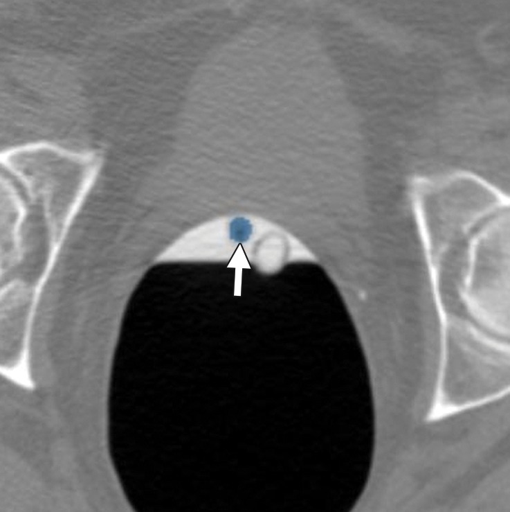
Mobile stool. (a) Magnified axial “flipped” 2D CT colonography image obtained in a prone position shows a submerged CAD polyp candidate (arrow) that is impossible to disregard without comparing it to the findings on supine images. Although it appears detached from the colon wall, all adjacent sections must be viewed to ascertain the absence of a stalk. (b) Magnified axial 2D CT colonography image obtained in the supine position shows the CAD polyp candidate (arrow), which is mobile to the dependent dorsal surface of the rectum. After correlating with the supine images, it is clear that this structure is mobile because it settles to the dependent region of the rectum in both the supine and prone series, a finding consistent with mobile stool. A mobile head of a pedunculated polyp must be excluded. In other potentially mobile colon segments, the possibility of bowel rotation should be excluded.